Photo Gallery
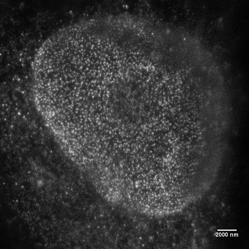
Superresolution Microscopy

Confocal image using SeTau-647
STED image using SeTau-647
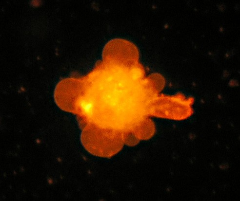
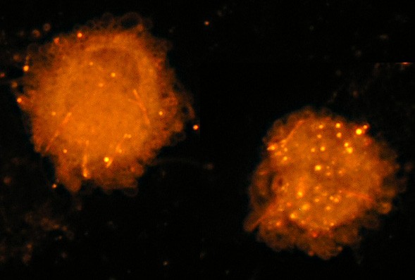
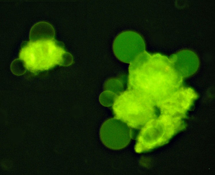
Saccharomyces Cerevisiae (Yeast) Cells

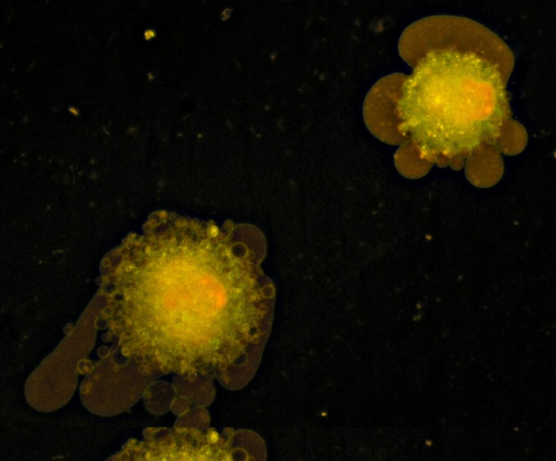
Saccharomyces cerevisiae cells stained with Square-460. Nectrotic cells with disrupted cell walls show bright yellowish-green fluorescence
Excitation: 470 nm
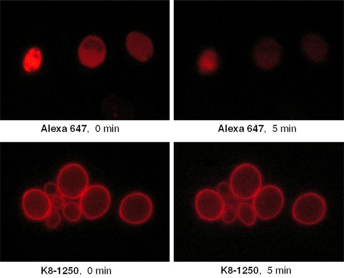
Saccharomyces cerevisiae cells stained with Alexa 647 and Square-635 at t = 0 and after 5 minutes of observation with an Olympus fluorescence microscope
Human Fibroblast Cells
Human fibroblast cells.
Excitation: 470 nm
Human fibroblast cells stained with 3-DAB. Excitation: 580 nm

Human fibroblast cells. Excitation: 470 nm
Human fibroblast cells stained with 2-DAB. Excitation: 580 nm
Human fibroblast cells stained with Square-680-Carboxy. Excitation: 580 nm
Human fibroblast cells stained with Square-670. Excitation: 580 nm
Human fibroblast cells stained with Square-655. Excitation: 580 nm
Human fibroblast cells stained with Square-460. Excitation: 470 nm
Erythrocytes

Human red blood cells (RBC, erythrocytes) stained with Square-460. Excitation: 470 nm
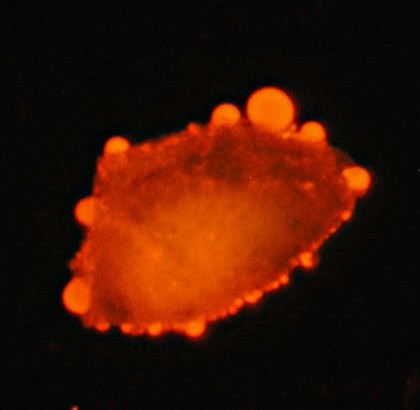
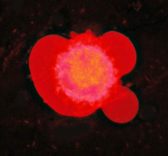
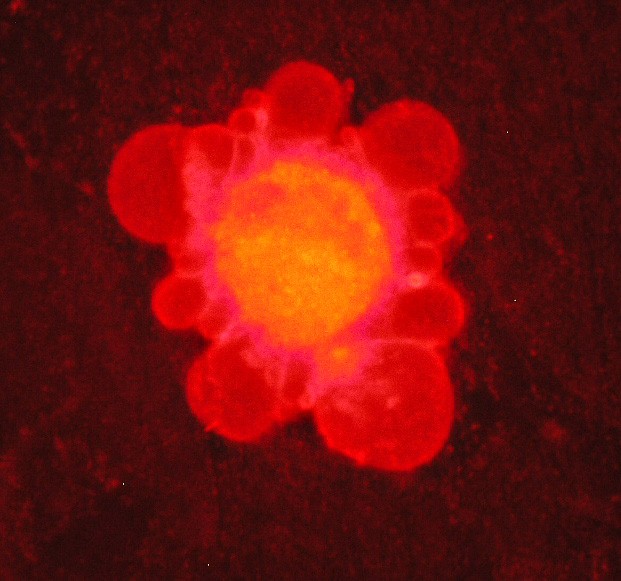
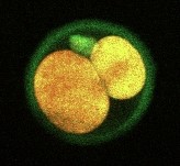
Mouse Egg Cells
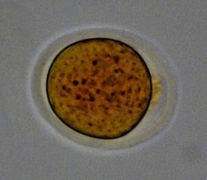
Mouse egg cell stained
with C8-4.
Excitation: 405 nm
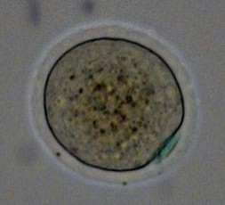
Mouse egg cell stained with Square-670-Carboxy (transmitted light mode)
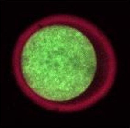
Mouse egg cell stained
with Square-670-Carboxy and C6-4
Mouse egg cell stained with Square-655 and Square-460